advertisement

Editors Selection IGR 11-1
Glaucoma staging system

Comment by Anders Heijl on:
13443 Categorizing the stage of glaucoma from pre-diagnosis to end-stage disease, Mills RP; Budenz DL; Lee PP et al., American Journal of Ophthalmology, 2006; 141: 24-30
See also comment(s) by Roger Hitchings •Mills et al. (278) have developed a new Glaucoma Staging System (GSS). This system has been used to analyze the costs of different stages of POAG, and the authors hope that it may provide an even greater service in providing a new standard for diagnosing, monitoring and cost-effectively treating the disease. When developing their new GSS, the authors reviewed some earlier staging systems, the Hodapp-Anderson-Parrish GSS, Brusini's GSS, Esterman's binocular system and the AGIS and CIGTS systems. The latter two were felt to involve more complex calculations, and the authors therefore decided to base their new system on the Bascom Palmer GSS. From this they constructed a system with six stages, where 0 represents no or minimal visual field defects, and stage 6 represents inability to perform Humphrey visual fields. Stages 1-4 represent early to moderate to advanced to severe visual field defects. The most important parameter is MD with cut of points -6 and-12 and -20 dB, but the authors apply modifiers in the form of additional criteria that would typically occur at that stage (see figure legend). If the patient fails to meet the additional criteria of his MD stage, he is categorized in the preceding (milder) stage. On the other hand, if he meets the MD criteria for his MD stage plus at least one of the additional criteria of the succeeding (worse) stage, the patient is categorized in that worse stage.
The new Glaucoma Staging System has advantages over earlier systems. It is, however, too complicated for extensive clinical use without computer. It is far from ideal for individual patient monitoringThe authors validated the system by retrospectively assigning unambiguous stages in Humphrey fields from 98 glaucoma charts. For staging purposes the new system has definite advantages over earlier standard systems. Firstly, this new GSS has more steps than previously described systems. Secondly, and probably more importantly, the authors have made an ambitious effort to add 'clinical severity' assessment to the classification by giving more weight to test outcomes closer to the point of fixation. This can certainly be of value for, e.g., cost analyses and epidemiology, and clearly represents - at least in some patients - a better description of the severity of the patient's disease than a simple MD value.
The main weakness of the new system is that the analyses are complicated and time-consuming, involving a large number of steps for each assessed patient or field (cf. figure legend). Certainly staging with the new GSS requires much fewer steps than that used in AGIS and CIGTS, but it is still too complicated as compared to Hodapp's classification, or a simple MD value, to be used clinically on a large scale without the availability of a computer program to assess the fields (plus of course the availability of the visual field data on the same computer). One may hope that such manipulation of perimetric data will become easier in the future.
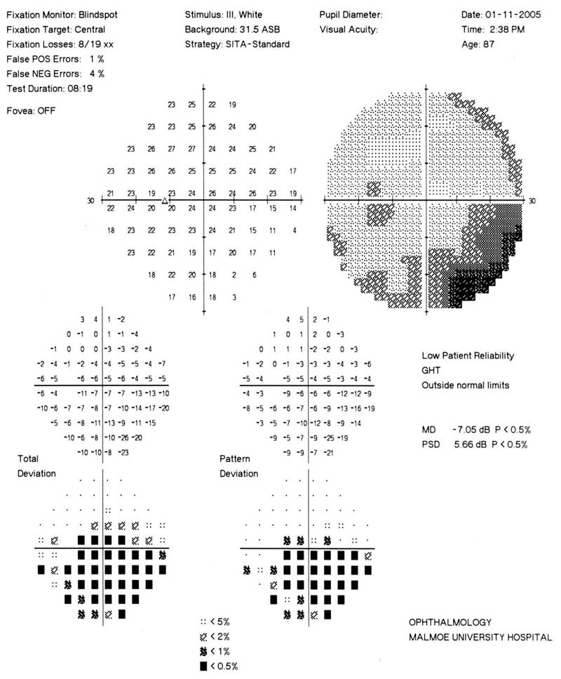
Example of staging of one eye (click on figure to enlarge):
The MD value (-7.05 dB) of this field is in the range of stage 2 moderate defects (MD of 6.01 to -12.00 dB). But in order to be classified as a stage 2 (moderate defect), one of the following three conditions also has to be fulfilled: A) on pattern deviation plot, greater than or equal to 25% but fewer than 50% all points depressed below the 5% level, and greater than or equal to 50 % but fewer than 25% of points depressed below 1% level; B) at least 1 point within 5° with sensitivity of < 15 dB but, no point within central 5° with sensitivity of < 0dB; C) only 1 hemifield containing a point with sensitivity <15 dB within 5° of fixation. Clearly none of these conditions are fulfilled and therefore the field will end up in stage 1 (early defect). If, on the other hand, any of the three conditions A-C above had been fulfilled, it would have been necessary to check where any of three conditions associated with stage 3 (advanced defect) have been fulfilled.
The authors suggest that the "GSS may be used to monitor the long time progression of disease in glaucoma patients," but warn that further validation is needed for this application. In my opinion a staging system like this is far from ideal for individual patient monitoring. The number of progression stages, even if larger than in previous GSSs, are fewer than those that can be obtained with ordinary visual field testing, and the described GSS like all other GSSs cannot help clinical management by giving a rate of progression. Certainly MD or Mean Sensitivity, Performance values or similar summary statistics have weaknesses in this regard (most importantly because they will be influenced by media opacities), but for disease monitoring purposes it is better to replace MD with an improved continuous variable index that could be used to calculate rate of progression, rather than to resort to staging.
Comments
The comment section on the IGR website is restricted to WGA#One members only. Please log-in through your WGA#One account to continue.Log-in through WGA#One


