advertisement


Optic Nerve Head Biomechanics
Claude F. Burgoyne, J. Crawford Downs, Anthony J. Bellezza
and Richard Hart
Use of Optic Nerve Head reconstruction to study the pathogenesis
of glaucoma.
Click here for representative,
rotating, 3-dimensional Optic Nerve Head reconstructions
Outline
- Basic Concepts
- IOP-related Stress and Strain are high in the ONH
- IOP-related axon loss is multi-factorial
- IOP-related connective tissue damage is multi-factorial
- 'Glaucomatous' is an appearance (not an etiology)
- The larger concept of IOP-related Optic Neuropathies
- Glaucomatous and 'Non-glaucomatous'
- How some non-IOP-related processes look 'glaucomatous
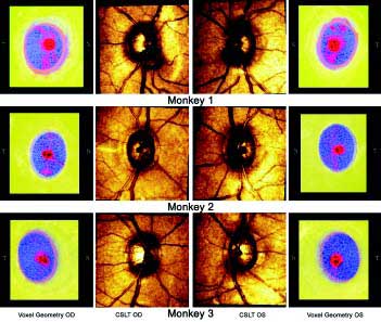
Fig. 1. 3D reconstructions of the Optic Nerve Head.
Summary
Burgoyne and colleagues have introduced the first digital 3D reconstructions
of the normal and early glaucoma monkey ONH connective tissues which demonstrate
that at the earliest stage of glaucomatous damage the connective tissues
of the lamina cribrosa and neural canal wall are profoundly and permanently
damaged. This damage should make the ONH more susceptible to further damage
at the same or even lower levels of IOP. Finite element models based on
3D reconstructions such as these will allow IOP-related stress and strain
within each portion of the connective tissue architecture to be estimated.
Such modeling will open the door to testing a series of hypotheses that
are central to a Biomechanical view of this neuropathy. These include:
- IOP-related connective tissue stress and strain are high at normal
levels of IOP;
- Regardless of the IOP at which they occur, IOP-related connective
tissue stress and strain govern the two central pathophysiologies of
the 'glaucomatous' optic neuropathies:
- IOP-related axonal damage which is multifactorial (compressive,
ischemic, glial cell mediated, and more�),
- IOP-related connective tissue damage - otherwise known as mechanical
failure - the predictable pattern of which is unique to each individual
ONH and centrally underlies the clinical phenomenon of glaucomatous
cupping.
*Figure originates from the paper 'Digital Three-Dimensional Reconstruction
of the Normal and Early-Glaucoma Monkey Optic Nerve Head Connective Tissue',
by Claude F. Burgoyne, J. Crawford Downs, Anthony J. Bellezza and Richard
Hart, which will appear in the December issue of IOVS. Reprinted with permission
of the publisher. The ONH as a Biomechanical Structure appears as an article
in the next issue of Progressive Retinal and Eye Research.
Issue 6-2
Change Issue
advertisement

|